Updated October 2022
Overview
EXTREMAG has five main user experiments, each with a variety of configurations to best suit user measurement requirements. In brief the following experiments are currently available, while more details are provided below.
Free space lab B2
- All-optical pump-probe Kerr measurements at variable temperature in a 10 T superconducting magnet.
- Time-resolved Kerr measurement of microwave driven magnetization dynamics (or all-optical pump-probe Kerr measurements) at variable temperature in a gas flow cryostat.
- Time-resolved scanning Kerr microscopy at room temperature with pulsed, microwave, or quasi-DC excitation of sub-micron materials and devices.
Microscopy lab B3
- All-optical pump, beam-scanning-probe ultrafast Kerr microscopy at variable temperature in a 5 T superconducting magnet.
- Wide field Kerr microscopy at variable temperature (heating or cooling) with all-optical pump, or electrical excitation of materials and devices.
All-optical pump-probe measurements at variable temperature
All-optical pump-probe experiments at variable temperature are available in three cryostats and use the tuneable Opera-F OPA output. Typically, two-colour measurements are performed using the 800 nm OPA signal output where 80% is used as the time-delayed ultrafast pump beam, while the remaining 20% is used for second harmonic generation of a low power 400 nm ultrafast probe beam. If alternative pump and probe wavelengths are required, please contact the SEO. The option for variable temperature is available using the following Oxford Instruments cryostats.
- Microstat He-R cryostat with rectangular tail and external electromagnet for moderate in-plane or out-of-plane field up to 0.5 T.
- Microstat MO superconducting magnet with up to 5 T out-of-plane field.
- Spectromag SM4000 superconducting magnet with up to 10 T out-of-plane field.
For all-optical pump-probe measurements at room temperature only, the Microstat He-R cryostat can be removed and replaced with a sample holder with 3-axis translation stage.
Microstat He-R cryostat
All-optical pump-probe
The Microstat He-R is located in the free space lab B2 so that it can be readily configured for two-colour, all-optical, pump-probe experiments. The wide optical access allows multiple samples to be mounted on the cold finger and the possibility to use a large angle of incidence to probe in-plane magnetized samples using the longitudinal Kerr effect. The pump is typically aligned at normal incidence. The 800 nm pump and 400 nm probe are weakly focussed with 200 mm and 100 mm focal length lenses to achieve a larger pump spot diameter and a more uniform excitation within the smaller region of the probe spot. A high turnover of samples is possible due to the rapid cool down time (~10 minutes) to the base temperature of ~4 K (likely higher on the cold finger). A heater on the heat exchanger can be used to stabilise intermediate temperatures and heat the sample to 300 K.
The Microstat He-R helium gas-flow cryostat with rectangular tail and optical access for all-optical pump-probe or optically detected FMR at variable temperature.
Optically detected ferromagnetic resonance
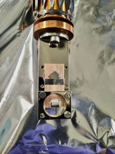
The Microstat He-R can also be used for time-resolved Kerr measurement of microwave driven ferromagnetic resonance (FMR) at variable temperature. The cryostat probe has an SMB RF feedthrough and coaxial cable to a coplanar waveguide (CPW) mounted on the rear side of the cryostat cold finger. The CPW has a 0.5 mm wide centre conductor on a ceramic filled, high thermal conductivity substrate (Arlon1000) onto which samples can be placed. Samples with substrate thickness <0.5 mm are mounted over the centre conductor of the CPW for excitation by the in-plane component of the microwave or pulsed magnetic field generated by the current waveform passing through the CPW. An externally applied magnetic field (up to 0.5 T) can be applied either in-plane along the length of the CPW, or out-of-plane as required.
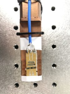
Coplanar waveguide and multiple samples on the cold finger of the Microstat He-R.
Magneto-resistance measurements
Variable temperature electrical measurements may also be possible on request via the RF feedthrough or original 10 pin DC sample connectors using a Keithley 6221/2182A (AC and DC Current Source/Nanovoltmeter) Delta Mode System.
Microstat MO 5 T superconducting magnet
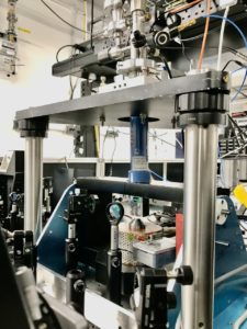
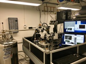
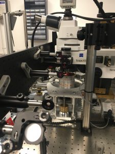
The Microstat MO (MO) is a helium gas flow superconducting magnet located in the microscopy lab B3. It is equipped with a two-colour, all-optical-pump, beam-scanning-probe microscope that has been constructed for ultrafast time-resolved imaging at low temperature and high magnetic field. The MO is mounted on its side so that its magnetic field lies perpendicular to the sample surface and parallel to the incident pump and probe beam in a polar Kerr effect geometry. The field direction is along the axis of the cylindrical body of the MO. The fixed position 800 nm pump and scanned 400 nm probe are focused using the same microscope objective lens (typically x5 or x20) such that optimum focus of the probe leads to a defocused pump with spot size observable within the maximum field of view, but significantly larger than the focused probe spot. Measurements can be performed at variable temperature from room temperature down to ~5 K, with magnetic field up to ~2.5 T. A heater on the heat exchanger can be used to stabilise intermediate temperatures and heat the sample to 300 K. Careful planning of user time with the MO system is required due to its ~5 hour cool down time.
The Microstat MO 5T superconducting magnet equipped with two-colour, all-optical-pump, beam-scanning-probe, polar Kerr microscopy.
Spectromag SM4000 10 T superconducting magnet
The Spectromag SM4000 (Spectromag) is located in the free space lab B2 and configured for two-colour, all-optical, pump-probe experiments in either reflection or transmission. The limited optical access allows only one sample within the field of view, but the sample probe can accommodate multiple (up to 4) samples, which can also be changed within ~1-2 hours when the system is cold. The time-delayed pump is typically aligned at normal incidence and the probe at ~10-15 degrees from normal. Flip mirrors in both optical paths allow the pump and probe paths to the readily swapped if needed. The 800 nm pump and 400 nm probe are weakly focussed with 400 mm and 200 mm focal length lenses to achieve larger pump spot diameter and a more uniform excitation within the smaller region of the probe spot. Two detectors are available for simultaneous measurement of the Faraday effect in transmission and polar Kerr effect in reflection.
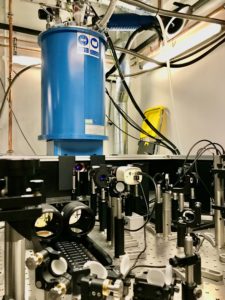
Two-colour, all-optical, pump-probe in the Spectromag SM4000.
The variable temperature insert (VTI) can be rapidly cooled (~10 mins) from an idle temperature of ~170 K by the flow of He gas (from the magnet He reservoir) directly over the sample. A heater on the sample probe and VTI can be used to stabilise intermediate temperatures and heat the sample to 300 K. Two-colour all-optical pump-probe has been demonstrated at an idle temperature of 170 K and up to 5 T, and at base temperature (<1.4 K) up to 1 T, while persistent mode of the magnet at 8 T has been demonstrated over ~12 hours. Careful planning of user time with the Spectromag is required due to its ~2 day cool down time once under high vacuum.
The Spectromag probe with two sample locations.
Time-resolved scanning Kerr microscopy
Time-resolved scanning Kerr microscopy (TRSKM) can be used to perform room temperature time-resolved measurements and imaging of magnetization dynamics with picosecond temporal resolution and spatial resolution down to 100s of nanometers.
The microscope is equipped with a piezoelectric scanning stage with 300 microns travel in x-, y-, and z-directions, and a quad-pass optical delay line for up to 8 ns of time delay. The microscope can use the 1040 nm, 80 MHz, 140 fs output of the Fidelity laser or its 520 nm second harmonic, or the signal output of the Opera-F OPA at 800 nm, 1 MHz, and <50 fs or its second harmonic at 400 nm. Electronic pulse generators operating at 80 MHz or 1 MHz may be used respectively for either 30 ps rise time, 70 ps duration, and ~5 V amplitude impulses for excitation of high frequency precession, or ~200 ps rise time, 2 ns, 30 V pulses for large amplitude dynamics and switching experiments. Alternatively, quasi-DC waveform excitation may be used, e.g. for spin orbit torque devices.
A small quad-pole electromagnet can be used sweep an in-plane magnetic field applied along any azimuthal direction (up to 50 mT). For larger static applied fields, a reconfigurable permanent magnet array (up to 100 mT in-plane and 300 mT out-of-plane) can be used for enhanced mechanical stability by eliminating the electromagnet and associated heating. The addition of a motorised translation stage and rotary mount will allow for automation of the field applied using the permanent magnet array.
The TRSKM can be used in three configurations for different sample types.
Continuous magnetic films
For continuous magnetic films on transparent substrates, the sample can be placed face down onto a separate coplanar waveguide (CPW). The CPW is designed for 50 Ohm characteristic impedance on a high frequency printed circuit board (Arlon 1000). Samples on sufficiently thin (<0.5 mm) opaque substrates can be placed face up, but the magnetic field excitation will be weaker due to the distance of the film from the CPW. Substrates should be insulating to avoid the shielding effects of eddy currents that doped substrates can present.
Micro- and nano-structured magnetic materials
For micro- and nano-structured magnetic materials an integrated CPW (or coplanar stripline) fabricated using photolithography is recommended. Such a waveguide can deliver a sizeable pulsed or microwave magnetic field for efficient excitation of micro- and nano-structured elements. At the same time, the integrated CPW serves as a location guide for small regions of patterned material, removing the need to fabricate large arrays. While patterned materials can be studied using the separate PCB CPW described above, precise alignment of sub-mm regions of patterned material with the centre conductor is challenging, while the excitation efficiency is compromised by the reduced proximity to the CPW, particularly on opaque substrates. Integrated waveguides can be designed with impedance matched large bond pads for contact with conductive paint, or with microscale contact pads to land high frequency microwave probes (preferred), or for wire bonding (not readily available). Microwave probes are available in a signal-ground (S-G), G-S, and G-S-G configuration with G-S pitch of 150 um or 300 um.
Magnetic devices
For devices, including spintronic, magnonic, magneto-acoustic, and magneto-electric, a pulsed, microwave or quasi-DC electrical waveform can be passed through the device itself to use the intended device mechanism for excitation, e.g. spin-transfer and spin-orbit torque, piezoelectric control of magnetostriction, or voltage-controlled magnetic anisotropy. Impedance matching of device contacts should be considered, if possible, particularly if the contact length is comparable to, or larger than the wavelength of the excitation waveform. Bond pads should be designed for use with microwave probes, as described above. The field of view in the TRSKM is small when measuring with the highest spatial resolution, and so nearby registration marks or numbers aid device identification.
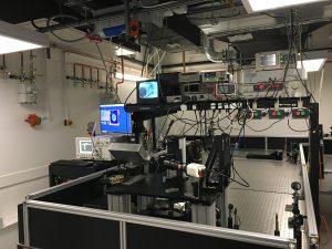
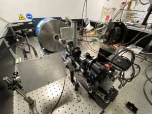
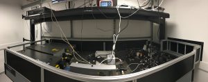
The time-resolved scanning Kerr microscope for room temperature imaging of picosecond magnetisation dynamics.
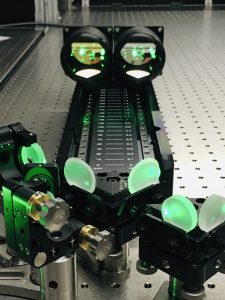
Quad-pass, 8 ns optical delay optics.
TRSKM can be performed with a weakly focusing 40 mm plano-convex lens for continuous film samples, or microscope objective lenses up to x60. For the highest spatial resolution combined with the use of microwave probes, a long working distance (~12 mm) high NA microscope objective is used to accommodate the probes beneath the objective, which can be used to fucus the probe laser directly between the S and G contacts of the microscale probes. In situ atomic force and magnetic force microscopy can be used in the TRSKM on request by adding a compact atomic force microscope to the microscope column allowing for characterisation of the sample topography and equilibrium magnetic state in an applied magnetic field (up to ~100 mT).
Electrical measurements may also be possible on request using a Keithley 6221/2182A (AC and DC Current Source/Nanovoltmeter) Delta Mode System.
THz spectroscopy and imaging
A THz spectrometer will be under construction from Autumn 2022 for use in the free space lab B2. The system will use fibre-coupled photoconductive antenna emitters and detectors for rapid integration with the either the Microstat He-R cryostat or the Spectromag 10 T magnet for variable temperature THz spectroscopy in an applied magnetic field.
The development of THz spectroscopy inside the laser enclosure. THz spectroscopy is now located in the Free Space Lab B2.
Wide field Kerr microscopy
High resolution wide field Kerr microscopy is available at room temperature, low temperature using the helium gas flow microscopy cryostat. In-plane or out-of-plane magnetic fields may be applied to samples in each case. The microscope can be equipped with a rotatable electromagnet for in-plane magnetic fields up to 1.3 T, or a polar electromagnet for out-of-plane magnetic fields up to 0.9 T. Low temperature microscopy down to below 10 K can be performed in an out-of-plane field up to ~100 mT, or an in-plane field up to ~700 mT. The microscope is sensitive to all three components of the sample magnetisation using longitudinal and polar Kerr effects. The microscope features piezoelectric drift correction for optimised differential imaging, and spatially resolved magnetometry (hysteresis loop measurement) from user defined regions of interest.
Electrical measurements may also be possible on request using a Keithley 6221/2182A (AC and DC Current Source/Nanovoltmeter) Delta Mode System. Note that piezoelectric drift correction is not possible for fixed electrical probe connections.
The high resolution wide field Kerr microscope with low temperature capability using a helium gas flow microscopy cryostat. A lower resolution overview wide field Kerr microscope is available for larger samples, or samples with large domain structure.
The system is also equipped with a lower resolution overview wide field Kerr microscope for larger samples or samples with large domains.
A pulsed laser optical pump with tuneable wavelength, or microscale electrical probes to devices are available for this instrument on request and subject to specific requirements.
In the longer term time-resolved wide field Kerr microscopy will be developed by using the femtosecond lasers as a light source for the wide field microscope.
The optical set up for optical pulse excitation of samples in the microscopy cryostat of the wide field Kerr microscope, equipped with polar field solenoid.